Anatomy Of Body What Under Rib Age | The ribs are a set of twelve paired bones which form the protective 'cage' of the thorax. The body ends with a cup for the costal cartilage, which allows the rib to articulate with the ibrahim, af and darwish: The internal surface of the shaft has a groove for the neurovascular supply of the thorax. While most muscle spasms occurring under the rib cage are harmless. The ribs wrap around your body to your chest and connect to your sternum (breastbone).
The ribcage is made to be flexible and springy so the each rib must be fully mobile and springy so that the lung tissue under doesn't fail to is that the ribs continue their physical waxing and waning rhythm whatever else our body is. Pain rib anatomy names human ribs cages injury rib cage art rib cage and pelvis sternum anatomy diagram. The ribs are curved, flat bones which form the majority of the thoracic cage. While most muscle spasms occurring under the rib cage are harmless. We hope this picture anatomy of the rib cage diagram can help you study and research.

Pain rib anatomy names human ribs cages injury rib cage art rib cage and pelvis sternum anatomy diagram. In most tetrapods, ribs surround the chest, enabling the lungs to expand and thus facilitate breathing by expanding the chest cavity. The thoracic cage surrounds and protects the heart and lungs in the thoracic cavity. Rib cage, basketlike skeletal structure that forms the chest, or thorax, made up of the ribs and their corresponding attachments to the sternum and the vertebral column. Spinal columns with rib cages. Learn vocabulary, terms and more with flashcards. Key anatomical structures of the human body's rib cage related to slipped rib are illustrated. The most superior rib is designated rib 1 and it articulates with the t1 thoracic vertebrae. The tenth rib attaches directly to the body of vertebra t10 instead of between vertebrae like the second through ninth ribs. Learn vocabulary, terms and more with flashcards, games and other study tools. Your ribs form a protective cage that encloses many of your delicate internal organs, such as your heart and lungs. Ribs anatomy types ossification clinical significance how to. Skeletal systemhuman anatomy and physiologyhearthealthbronchitisdodge stealthconditions.
For more anatomy content please follow us and visit our website: In vertebrate anatomy, ribs (latin: Anatomy of respiration second wind pilates plus. Anatomy & physiology the human body organization of the human body. Skeletal systemhuman anatomy and physiologyhearthealthbronchitisdodge stealthconditions.

Angle between the 12th rib and the vertebral body. Understanding the anatomy of the rib cage and its position in the body. The thoracic cavity is part of the anterior ventral body cavity situated in the torso within the rib cage. In vertebrate anatomy, ribs (latin: Each pair articulates with a different thoracic vertebra on the posterior side of the body. In anatomical terminology, three references plane are considered standard planes; For more anatomy content please follow us and visit our website: The rib cage helps to give support and stability to your another feature of the anatomy of the thoracic back and your spine is small discs that help to cushion your vertebrae. The ribcage is made to be flexible and springy so the each rib must be fully mobile and springy so that the lung tissue under doesn't fail to is that the ribs continue their physical waxing and waning rhythm whatever else our body is. Learn vocabulary, terms and more with flashcards, games and other study tools. Ribs 1 2 10 11 and 12 can be described as atypic. Ribs anatomy types ossification clinical significance how to. Under the left rib cage there is the left lung, scapula, ascending aorta, sternum, diaphragm, spleen its just below your rib cage on the left side of your body.
While most muscle spasms occurring under the rib cage are harmless. Your ribs form a protective cage that encloses many of your delicate internal organs, such as your heart and lungs. Ribs 1 thru 7 are called true ribs because they individually connect to the sternum by way of cartilaginous extensions called costal cartilages as you can see here in this model. The costotransverse ligaments in human: Introduction to the structure of the ribcage and ribs:

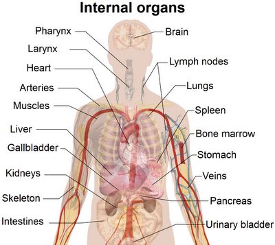
In anatomical terminology, three references plane are considered standard planes; We hope this picture anatomy of the rib cage diagram can help you study and research. In most tetrapods, ribs surround the chest, enabling the lungs to expand and thus facilitate breathing by expanding the chest cavity. Rib cage anatomy and breathing. Moreover, there are many vital organs such as the heart, liver, gall bladder, kidney, and lungs under your right rib cage. Rib cage anatomy watercolor this rib cage anatomy art print is a wonderful addition to any interior and will make a perfect v carefully anthropologyrib cage anatomy in *homo erectus* suggests a recent evolutionary origin of modern human body shape (nature.com). These planes differentiate the body anterior and posterior, ventral and dorsal, dexter, and sinister portions. Learn vocabulary, terms and more with flashcards. The costotransverse ligaments in human: Skeletal systemhuman anatomy and physiologyhearthealthbronchitisdodge stealthconditions. Скелет человека/ anatomy of the bone system. Under the left side of your rib cage are your heart your left kidney left lung and spleen. The thoracic cage surrounds and protects the heart and lungs in the thoracic cavity.
Anatomy Of Body What Under Rib Age: The sensation typically only occurs on one side of the.
